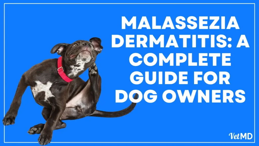
Malassezia dermatitis or Yeast dermatitis in dogs is commonly seen fungal infections in pets which is primarily caused by fungus of Malassezia group. Malassezia has long been recognized as a common perpetuating factor in otitis externa. A significant development in veterinary dermatology in recent years is the recognition that Malassezia can be linked to generalized dermatitis, and that antifungal treatment can lead to substantial improvements in these cases.
The Malassezia organism
Species and strains Malassezia is a genus of commensal yeasts found on mammalian and avian skin. Malassezia pachydermatis uniquely does not require lipid supplements during culture in vitro. There are six lipid-dependent species: M. furfur, M. sympodialis, M. globosa, , M. obtusa, M. restricta and M. slooffiae.
The lipid dependent species are most commonly isolated from humans. In contrast, M. pachydermatis can be recovered from a variety of animals, especially carnivores. So far only M. pachydermatis has been isolated from dogs, but M. sympodialis and M.globosa, as well as M. pachydermatis, have been isolated from cats. Seven strains of M. pachydermatis have been identified. None has been associated with virulence, although type Id was restricted to dogs, whilst others were more ubiquitous.
(a) Morphology
Malassezia is a single-cell yeast with a thick cell wall. Individual cells are ovoid to globular or cylindrical. Cells in the process of budding form the characteristic ‘peanut’ to ‘Russian doll’ shape.
(b) Ecology
Malassezia can be recovered from 3-day-old puppies, suggesting there is early maternal transfer by licking, grooming or from the vagina. Malassezia is commonly found in the ear canals, anal sacs, interdigital skin and mucocutaneous junctions (lips, prepuce, vagina, anus) of healthy animals. It is rarely recovered from skin elsewhere on the body. Mucosal sites in particular may be reservoirs from which yeasts are seeded onto the skin by licking and grooming. Evidence for this is seen in Basset Hounds, where successful topical treatment is associated with a reduction of both mucosal and cutaneous Malassezia populations.
Malassezia yeasts colonize the superficial layers of the epidermal and infundibular stratum corneum. They appear to have a symbiotic relationship with cutaneous staphylococci, producing both mutually beneficial growth factors and a favourable microenvironment. Concurrent pyoderma frequently complicates Malassezia dermatitis. Furthermore, treatment regimes that also reduce bacterial numbers are superior to those with antifungal action only.
(c) Virulence factors and host susceptibility
Precisely how a commensal organism becomes a pathogen is unclear: the balance with host defences must shift in favour of the microorganism. Malassezia can have a significant pathogenic role when a combination of virulence factors and microclimate allows the yeasts to overwhelm the physical, chemical and immunological host defences that normally limit colonization and proliferation.

i. Virulence factors
M. pachydermatis expresses a variety of protein or glycoprotein adhesion molecules that bind to carbohydrate ligands on canine corneocytes. However, neither the expression of adhesion molecules nor adhesion to corneocytes in vitro has been associated with virulence in Malassezia dermatitis. Similarly, the ability of M. furfur to adhere to human keratinocytes is not linked to seborrheic dermatitis.
Malassezia yeasts secrete various proteases, lipases, phospholipases, lipoxygenases and other enzymes, which cause proteolysis, lipolysis, alter cutaneous pH, activate complement and trigger the release of inflammatory mediators. These changes could alter the cutaneous microclimate in favour of Malassezia and staphylococci, as well as contributing to inflammation and pruritus. Despite this, no specific link with virulence has been demonstrated.
ii. Host factors
The lack of an association between biological activity and virulence suggests that Malassezia is an opportunistic pathogen, able to establish wherever there is a permissive microenvironment.
iii. Anatomy
Anatomical features (body folds, pendulous lips, hairy feet), inflammation, exudates and licking can create a warm, moist microenvironment. Keratinization defects and endocrine disorders may increase humidity, and alter the quantity and quality of sebum, although the relationship between sebum production and Malassezia growth is unclear. Additionally, damage to the stratum corneum caused by self-inflicted trauma, keratinization issues, or metabolic disorders may create an environment where Malassezia can thrive.
iv. Breed susceptibility
Certain breeds are predisposed to Malassezia dermatitis. The nature of the predilection is unclear. It could include anatomical features, specific host factors and predisposition to diseases with secondary Malassezia involvement. Mucosal and cutaneous Malassezia populations are elevated in healthy Basset Hounds, suggesting factors that facilitate Malassezia colonization play a role in the susceptibility of this breed.

Breeds predisposed to Malassezia dermatitis
| Basset Hounds |
| West Highland White Terriers |
| Cocker Spaniels |
| Shih Tzus |
| Dachshunds |
| Miniature Poodles |
| German Shepherd Dogs |
| Australian Silky Terriers |
| English Setters |
v. Immune response
Malassezia does not invade the stratum corneum. Even so, immune responses to Malassezia organisms can be detected in healthy and affected dogs. At least 14 different protein antigens have been identified. Dogs with Malassezia dermatitis tend to recognize more antigens than healthy dogs, but no association between the pattern of antigen recognition and any particular Malassezia strain or virulence has been demonstrated.
Affected dogs also have elevated serum IgA and IgG titres compared to healthy dogs, but this does not appear to be protective. Basset Hounds with Malassezia dermatitis exhibit decreased lymphocyte responses compared to healthy dogs, suggesting that protective immunity is associated with strong cell- mediated rather than humoral responses. Positive delayed skin test reactions, however, are seen in both healthy and affected Bassets.
A significant proportion of human patients with atopic dermatitis develop a hypersensitivity response to Malassezia. Malassezia dermatitis and otitis also frequently complicate canine atopic dermatitis. Atopic dogs have larger populations of Malassezia at mucosal and interdigital sites than healthy dogs. Furthermore, intradermal test reactivity to a crude Malassezia extract was seen in atopic, but not healthy, dogs. In contrast, immediate skin test reactivity in non-atopic Basset Hounds with Malassezia dermatitis is infrequent. Recent studies have also detected higher levels of Malassezia specific IgG and IgE in atopic dogs than in healthy dogs. This suggests that Malassezia could participate in atopic dermatitis by acting as allergens or superantigens, although there is no evidence of this as yet.
vi. Other predisposing factors
Whether Malassezia is a primary disease or a secondary complication is controversial. Malassezia has been associated with a number of primary conditions in dogs and cats, but other studies showed that dogs with atopy, keratinization defects and endocrinopathies were no more at risk of Malassezia dermatitis than dogs with any other dermatological problem. Treatment with glucocorticoids or antibiotics does not appear to be a factor. However, in the author’s experience, primary Malassezia dermatitis is uncommon in most breeds, and is most frequently secondary to an underlying hypersensitivity.
Possible underlying conditions in Malassezia dermatitis.
| Body folds |
| Scabies |
| Demodicosis |
| Atopic dermatitis |
| Adverse food reaction |
| Endocrinopathies |
| Keratinization defects |
| Superficial necrolytic dermatitis/necrolytic migratory/erythema/hepatocutaneous syndrome |
| Zinc responsive dermatosis |
| Feline paraneoplastic syndrome |
| Feline thymoma |
| Feline leukaemia virus/feline immunodeficiency virus |
| Feline acne/facial dermatitis |
| Immunosuppressive therapy |
| Psychological stress |
(d) Zoonotic potential
Malassezia pachydermatis can transiently colonize humans. The likely cause of M. pachydermatis-associated septicemia, meningitis, and urinary tract infections in an intensive care nursery was the colonization of staff members’ hands by pet dogs. The yeast then persisted through patient-to patient transmission. The elderly, AIDS sufferers or patients undergoing chemotherapy may also be at risk. This underlies the need to observe hygienic precautions when handling healthy animals, as well as those affected by Malassezia dermatitis.
What are the Clinical signs of Malassezia Dermatitis?
(a) Symptoms of Malassezia Dermatitis in Dogs
Malassezia dermatitis can occur in any breed, although certain breeds are predisposed. There does not appear to be any age or sex predilection. The major clinical sign is pruritus, which can be severe, causing frenzied scratching that can be misinterpreted as a neurological problem. In the early stages there is erythema and greasy exudation, scaling and crusting. Chronic Malassezia dermatitis is characterized by greasy alopecia, lichenification and hyperpigmentation. Clinical signs can be focal or generalized, diffuse or well demarcated. Commonly affected sites include the ears, lips, muzzle, feet, ventral neck, axillae, ventral body, medial limbs, perianal skin and tail. Affected dogs often have a rancid, musty or yeasty odour.

Less commonly dogs present with recurrent interdigital furunculosis or ‘cysts’. Malassezia can also cause paronychia with a waxy exudate and brownish discoloration of the nails. Malassezia is also a rare cause of stomatitis, pharyngitis and tonsillitis.

An underlying condition and/or the staphylococcal pyoderma present in many cases often complicate the clinical picture.
(b) Symptoms of Malassezia Dermatitis in Cats
Malassezia dermatitis is less common in cats than in dogs, but has been associated with otitis externa. Pruritus is a less constant feature than in dogs. Malassezia can be involved in recalcitrant feline acne and facial dermatitis characterized by erythema and comedones, with large dark tightly adherent scales and follicular casts. Other cats may present with generalized scaling and erythema. Some cats, particularly Devon Rex, present with paronychia or waxy, red-brown discoloration of the nails. Generalized erythema and greasy scaling has been associated with Malassezia dermatitis in cats with thymoma and paraneoplastic alopecia.


How Malassezia Dermatitis is Diagnosed?
The differential diagnosis list is extensive and complicated by the fact that many conditions are risk factors for secondary Malassezia dermatitis.
Differential diagnoses for Malassezia dermatitis
| Fleas |
| Scabies |
| Demodicosis |
| Dermatophytosis |
| Staphylococcal pyoderma |
| Atopic dermatitis |
| Adverse food reaction |
| Drug reactions |
| Contact dermatitis |
| Seborrhoea oleosa |
| Seborrheic dermatitis |
| Feline acne/facial dermatitis |
| Acanthosis nigricans |
| Epitheliotropic lymphoma |
Essentially, Malassezia should be considered in any pruritic dermatitis, particularly if associated with:
- Erythema
- Scaling
- Greasy or waxy exudate
- Hyperpigmentation
- Lichenification.
However, identification of Malassezia organisms does not preclude the possibility of an underlying condition, nor the need for further diagnostic steps. No agreed diagnostic criteria have been established. Most authors consider demonstration of elevated numbers of organisms, and a good clinical and mycological response to antifungal treatment are diagnostic.
(a) Cytology
Cytology is quick, easy, cheap and non-invasive. Direct impression on to a glass slide is possible on accessible skin; it is helpful if the skin is very moist or waxy. Blunt scalpel blades or cotton swabs can be used to collect waxy exudate from the ears, nail folds, body folds and feet. Tape strips are effective, unless the skin is very moist or inaccessible. However, practice and proficiency, rather than technique, is most important.
Slides should be gently heat-fixed (alcohol would remove many of the organisms) and then stained in a modified Wright’s stain (such as Diff-Quik®) or methylene blue. The staining solutions should be changed regularly, as yeasts can collect in them leading to false positive diagnoses. Tape strips need not be fixed before staining. Experimenting with locally available adhesive tape may be necessary to find one that resists the stain used.
Slides are first examined under low magnification (X40-X100) to assess the quality of staining and identify regions with abundant squames for detailed analysis. Malassezia appear as small oval to peanut or snowman shapes, often forming rafts on the surface of squames. They most frequently stain blue-purple, but can appear red-pink or pale blue. Some Malassezia fail to stain, but their refractile cell wall can be picked out with a closed condenser. Using the oil immersion lens (X1000) is the most accurate way to find Malassezia, but with practice they can be easily identified using the dry lens (X400).

There is no standard accepted number of organisms needed to diagnose Malassezia dermatitis. Estimates of Malassezia populations on healthy skin range from <8 yeasts/cm2 to <1 yeast per high power field (X400). Estimates of clinically significant Malassezia numbers range from >2 yeasts per high power field to > 10 yeasts per oil immersion field (X1000).
It is likely that these figures reflect differences in technique, breed and body sites. Furthermore, Malassezia are often found in rafts associated with squames and may not be uniformly distributed across a slide. Hence, an individual’s clinical experience and acumen is as important as relying on numbers. The author currently uses a benchmark of ≥5 yeasts per high power field . In practice, only occasional Malassezia yeasts are found on healthy skin.
(b) Culture
Malassezia pachydermatis will grow on Sabouraud’s medium, although the lipid-dependent species require supplemented media, such as modified Dixon’s agar. Samples can be collected by swab or direct contact for quantitative and semi-quantitative culture, although this requires an incubator and supply of sterile media. Plates should be placed in contact with the skin for 5-1 a seconds, then cultured at 32-37°C for 3-7 days. Colonies are small, cream to yellow, dome shaped, smooth to slightly wrinkled, with a regular to slightly lobed edge. However, as Malassezia are commensal organisms, isolation is not necessarily significant. Typically, <1 colony forming unit can be isolated from healthy canine skin, but much higher populations can be isolated from the lips and interdigital skin.
(c) Skin biopsy
Malassezia can be present in the overlying keratin crust and hair follicles, but organisms are often removed by processing. Malassezia can also be an incidental finding in biopsy samples from other dermatoses. The histopathology of Malassezia dermatitis is characterized by acanthosis, hyperkeratosis and a superficial inflammatory infiltrate.
What is the Treatment of Malassezia Dermatitis
Several topical and systemic treatment options are available to treat Malassezia dermatitis. Treatment should be tailored to the individual case.
(a) Topical therapy
Topical therapy is generally the most cost-effective and safest treatment. However, it is also the most labour intensive, and therefore not necessarily the most appropriate in all cases. Topical antifungal products include:
- 2% miconazole and chlorhexidine shampoo
- 1% selenium sulphide shampoo
- 1-4% chlorhexidine scrubs
- Enilconazole rinse
Localized areas of Malassezia dermatitis (e.g. body folds) can be treated with focal application of an antifungal product, but the whole body should be treated in multifocal or generalized Malassezia. It is particularly important to treat the ears, mucocutaneous junctions and feet, as these are likely reservoirs of Malassezia. Treatment should be continued daily to three times weekly until resolution, then as necessary to maintain the improvement. Treatment with degreasing shampoos or antibacterial products may also be necessary initially. Adverse reactions are uncommon, although most of the antifungal products can be drying and irritating, and may need to be combined with emollient rinses or shampoos.
Other treatment options include imidazole-containing shampoos, lotions, ointments and creams licensed for medical use (e.g. ketoconazole, clotrimazole etc.). These are not licensed for use in animals in the UK and therefore should only be used if necessary, but the creams can be useful for treating focal lesions. Terbinafine lotion (1 %) is effective in human seborrheic dermatitis.
(b) Systemic therapy
Where topical therapy is impractical or ineffective, systemic triazole antifungals can be used:
- Ketoconazole @ 2.5-10 mg/kg orally bid
- Itraconazole @ 5-10 mg/kg orally sid
However, these drugs are expensive, not licensed for animals, and can have side effects. Griseofulvin is not effective against Malassezia. Clinical improvement should be obvious after 7-14 days, although treatment should be continued for 7-14 days beyond clinical cure. Maintenance doses 2-3 times weekly may be necessary in some cases. Systemic or topical antibacterial therapy may also be necessary.
Side effects can include anorexia, vomiting, diarrhoea and liver damage. Ketoconazole can also be teratogenic. Haematology and serum biochemistry need to be monitored during therapy.
What is the Prognosis of Malassezia Dermatitis?
The prognosis with either systemic or topical treatment is very good in most cases. However, unless an underlying cause is diagnosed and treated, it is likely that lifelong maintenance therapy will be necessary. A greater understanding of Malassezia dermatitis may allow us to explore targeted treatment of the mucosal reservoir population, immunotherapy or colonization with non-pathogenic strains in the future.


Pingback: Otitis Externa in Pets: Complete Guide from Signs to Cure
Pingback: Ringworm in Dog and Cat: Protect Your Pet From This Fungus -
Pingback: Canine Demodicosis (Demodex Mites Infection in Dogs) - VetMD
Pingback: Oatmeal Baths for Dogs: Everything You Need to Know About
Pingback: Anal Sac Disease in Dogs: Causes, Signs and Treatment Option
Pingback: Atopic Dermatitis: Causes, Signs and Treatment for Your Pet
Pingback: Golden Retriever: Everything You Need to Know About - VetMD
Pingback: Flea Prevention for Dogs: How to Safeguard Your from Fleas
Pingback: Pruritus in Pets: Understand the Cause, Signs and Treatment
Pingback: Understanding Why Your Dog’s Ears Smell and How to Help
Pingback: Food Poisoning in Dogs: Can Dog Get Food Poisoning?
Pingback: Dog Skin Allergies vs. Bug Bites: How to Tell the Difference
Pingback: Dog Food Allergies: What’s The Best Dog Food For Allergies
Pingback: Safe Over-the-Counter Medications for Dogs: A Quick Guide
Pingback: Skin Infections in Dogs: How to Identify and Treat Them
Pingback: Dog Shampoo Advice: Why Human Shampoo Isn’t Good for Dogs
Pingback: Pneumonia in Dogs: Causes, Symptoms, Diagnosis and Treatment