
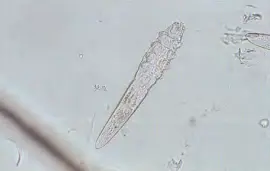
Canine demodicosis is a skin disorder associated with higher than normal populations of demodicid mites. Demodex canis, a slender mite with short stubby legs and a long tapering abdomen, is host-specific and apparently a normal inhabitant of canine skin. It lives mainly within hair follicles (rarely in sebaceous glands) and feeds on cells, sebum and epidermal debris. Adult mites measure 250-300 μm x 40 μm. Both a shorter (90-148 μm long) and a longer (334-368 μm long) demodicid mite have been found in some dogs. These represent either separate species, previously unrecognized, or mutant/aberrant forms of Demodex canis. The short variety is believed to live only on the skin surface whereas the longer mite has been found in pilosebaceous units.
Predisposing factors
Purebred dogs seem to have a higher risk of developing demodicosis compared to mixed breeds. One North American study found a predisposition to generalized demodicosis in the following breeds: Shar Pei, West Highland White Terrier, Scottish Terrier, English Bulldog, Boston Terrier, Great Dane, Weimaraner, Airedale Terrier, and Afghan Hound. However, there may be geographical variation in breed susceptibility. The tendency to develop demodicosis appears to be hereditary, and limited data suggest an autosomal recessive mode of transmission . Not breeding from clinical cases, or any of their siblings and parents, has been found to reduce or even eliminate the incidence of the disease in some lines.
Life cycle of Demodex Mites
The complete life cycle takes place on the skin. From lemon-shaped eggs, hatch larvae (six legs) which moult first into nymphs (eight legs) and subsequently into adults (eight legs).
Transmission of Demodex Mites
The only means of demodex mite transfer appears to be from bitch to sucking puppy by direct contact during the first few days of the pup’s life. Mites have been found in hair follicles from 16 hours after birth, appearing first on the muzzle. Mites are not found on stillborn puppies nor on puppies born by Caesarean section and reared away from the bitch. There is no evidence of horizontal spread between older dogs.
Pathogenesis
It is unknown why, if the mite is a normal inhabitant of canine skin and hair follicles, it should cause skin disease in some dogs and not others. It has been suggested that virulence may vary with the strain of mite. However, within a litter of puppies exposed presumably to the same strain of mite, some individuals may have severe clinical disease whilst others remain asymptomatic.
The discovery that clinical demodicosis could be induced by giving dogs antilymphocyte serum led to the theory that the condition might be linked to immunodeficiency. Further evidence for this comes from adult dogs developing demodicosis in association with severe neoplastic or metabolic disorders, or when treated with immunosuppressive drugs. However, if puppies with generalized demodicosis were severely immunocompromised they would be expected to develop widespread systemic infections and this does not happen. Equally, most adults with debilitating diseases or given immunosuppressive drugs do not develop demodicosis.
Some studies have demonstrated immunosuppression . Initially, this was thought to be induced by the mites themselves although later studies suggested that immunosuppression was a consequence of pyoderma (a common complication of demodicosis). More recent work has shown that the immune system of dogs with demodicosis becomes more compromised with both greater populations of mites and pyoderma. However, immunological abnormalities persist following resolution of demodicosis and pyoderma, and immunosuppression is not a precondition for the development of clinical demodicosis.
A hereditary, Demodex canis-specific, T-Lymphocyte defect of varying severity may be involved. Changes in hormone levels may also encourage mite proliferation: clinical demodicosis frequently develops around 6-9 months of age, a time when many puppies are becoming sexually mature.
Clinical Signs of Demodicosis in Dogs
Canine demodicosis is seen mostly in pure-bred dogs under 1 year of age. It may be localized or generalized. When feet are involved, the condition is known as pododemodicosis.
(i) Localized demodicosis
Localized demodicosis develops typically at 3-6 months of age. It is a mild disease, resolving spontaneously in 90% of cases , usually within 6-8 weeks, although lesions may wax and wane for months. Hair loss and erythema are often the presenting signs and owners may see patches of red, scaly skin, which tend not to be itchy. The face, especially around the eyes and mouth, is the most common site affected, followed by the forelegs. Hindlegs, trunk and ears are less frequently involved. Rarely, the condition may be restricted to the ear canals, producing a ceruminous, sometimes pruritic, otitis externa. It is very unusual for localized demodicosis to become generalized.
(ii) Generalized demodicosis
The generalized form of the disease usually starts in the first 18 months of a puppy’s life (juvenile-onset). Occasionally, it occurs de novo in older dogs (adult-onset). There is no universally recognized standard as to what constitutes ‘generalized’. It has been suggested that fewer than six lesions indicate localized disease and 12 or more lesions indicate the generalized condition, but each patient clearly needs to be assessed individually.
Juvenile-onset: Typical age of onset is between 3 and 18 months. Multiple poorly circumscribed areas of erythema, scaling , crusting, hair loss and hyperpigmented skin may be seen initially. Secondary pyoderma (usually associated with staphylococcal infection; less commonly Pseudomonas and Proteus) is common and leads to oedema, exudation and thick crust formation. Otitis externa may be present. Some dogs have nodules and other atypical lesions. English Bulldogs are reported to be predisposed to demodectic nodule development. Lesions are not normally itchy in the absence of pyoderma. Dogs are frequently depressed and may have peripheral lymphadenopathy. In dogs under 1-year old, generalized demodicosis resolves spontaneously in more than 50% of cases.
Adult-onset: This rare form of the disease occurs in dogs aged 4 years or more with no prior history of demodicosis. It follows a reduction in the dog’s resistance to the mites, which have previously been tolerated and kept in check by a healthy immune system. The condition is often associated with systemic disease (e.g. neoplasia, Cushing’s disease, hypothyroidism) or treatment with immunosuppressive drugs. Where associated with an internal disorder, demodicosis may be diagnosed before signs of that disorder are detectable. Atopic dogs with a long history of steroid therapy may sometimes develop adult-onset demodicosis.
Clinical signs are similar to those of juvenile-onset demodicosis. Severity is variable and the prognosis poor, particularly in cases where the underlying disorder cannot be corrected.
(iii) Pododemodicosis
A dog’s paws may form part of a generalized presentation or they may be the only region affected. Pedal lesions are particularly susceptible to secondary pyoderma and can be so painful as to cause severe lameness. Pododemodicosis can be very resistant to therapy, especially if lesions are infected with Pseudomonas. Old English Sheepdogs seem to be more prone than other breeds.
Diagnosis
A consideration of the history and clinical signs is important but to confirm the diagnosis, mites must be found. The most useful procedure for demonstrating demodicid mites is the deep skin scraping. Affected skin should be squeezed firmly to extrude mites from hair follicles. Hair plucks are also very useful, especially from areas that are difficult to scrape such as feet and face. Finding one adult mite may not be significant but if many adults or immature stages (eggs, larvae or nymphs) are found , the disease is confirmed. If in doubt, more scrapings from several parts of the body should be taken. In some cases, particularly with pedal lesions, skin biopsy specimens should be submitted for histopathology before the condition can be ruled out.
Differential diagnosis includes dermatophytosis, bacterial folliculitis and pemphigus foliaceus.
In adult-onset demodicosis, an underlying condition should always be sought through:
- Full haematology and biochemical profiles
- Thyroid evaluation (e.g. T4, TSH)
- Adrenal evaluation (e.g. dexamethasone suppression test)
- Chest and abdominal radiographs
- Urinalysis
- Heartworm serology (in countries where it is prevalent)
However, an underlying disorder may sometimes only become apparent months or even years later.
Treatment of Demodicosis in Dogs
The golden rule is always to avoid steroids no matter how tempting it is to use them. They may suppress an already compromised immune system and are contraindicated in all forms of demodicosis. In adult-onset demodicosis, response to any therapy may be incomplete unless predisposing factors have been addressed.
(i) Localized demodicosis
The condition in most young dogs does not require specific acaricidal treatment because of the likelihood of spontaneous resolution. Therapy may be completely unnecessary but washing with benzoyl peroxide, ethyl lactate or chlorhexidine shampoos can be helpful. Glucocorticoids may further suppress a compromised immune system and can lead to the condition becoming generalized. They should be avoided. In the USA, rotenone ointment and shampoo are licensed for treatment of localized disease.
(ii) Generalized demodicosis (including pododemodicosis)
Although the condition in many young dogs may resolve spontaneously, cases of generalized demodicosis should be treated with a suitable acaricidal product. Every effort should be made to ensure that dogs with generalized demodicosis are in optimal general health. A good diet should be given and suitable worming procedures performed regularly.
Owners should be advised to have their bitches spayed. There are two reasons for this: a bitch with a history of generalized demodicosis should not be used for breeding ; and intact females may relapse or the disease become refractory to treatment when the bitch is in season. Entire male dogs should be castrated to prevent breeding.
In adult-onset demodicosis, any underlying systemic condition should be corrected if possible.
All forms of glucocorticoids should be avoided to prevent further immune system suppression . Immunostimulants such as vitamin E and levamisole have not been found to improve the cure rate of generalized demodicosis.
Treatment of generalized demodicosis can be particularly troublesome in dogs with concurrent allergic skin disease for whom only glucocorticoids provide relief. Euthanasia is sometimes indicated for dogs that fail to improve with available treatments.
Amitraz: This is the only product licensed for treating canine demodicosis in the UK and is the treatment of choice. A 0.05% aqueous solution of amitraz (500 ppm) is applied, all over, at an initial frequency of once weekly. This strength of solution is obtained by diluting 50 ml amitraz into 5 litres of water.
In the USA amitraz is licensed for use as an 0.025% solution (250 ppm) to be applied every other week. Although frequency of application can often be reduced to every 2 weeks once the condition appears to be under control clinically, therapy should be continued until at least 1 month after scrapings have repeatedly failed to show any stages of mite, dead or alive. As an aid to monitoring the effects of therapy, skin scrapings and hair plucks should be carried out every 2-4 weeks.
Dogs with medium or long coats should be clipped to allow the solution better skin contact and greater penetration into hair follicles. Before amitraz is applied, dogs should ideally be washed to aid removal of scale, crust and exudate. For this purpose, one of the shampoos recommended above for treating localized demodicosis can be used. These shampoos may also exert antibacterial and follicular flushing activity. Appropriate systemic antibiotics are always indicated when pyoderma is present.
Amitraz should be avoided in dogs experiencing heat stress, as well as in pregnant or nursing females, and puppies under 3 months of age. In Chihuahuas, amitraz has been associated with a small number of deaths for which no cause could be determined, and use of amitraz in this breed is therefore contraindicated. There are no contraindications in other small or toy breeds. Some dogs show transient erythema, sedation or vomiting following amitraz application. Lethargy and vomiting are less likely to occur if dogs are prevented from licking themselves until the product has dried.
Side effects are likely to resolve spontaneously within 24-48 hours but atipamezole has been found to reverse the sedative effects of amitraz. In some countries, yohimbine (unavailable in the UK) is used to counteract amitraz toxicity.
Amitraz is a monoamine oxidase inhibitor (MAOI) and adverse reactions have been reported in dogs and people taking other MAOls (e.g. certain antihistamines, antidepressants and antihypertensive agents) following application of amitraz to the dog. Nausea and dizziness may occur following inhalation. Amitraz can induce hyperglycaemia and should not be used in diabetic dogs nor applied in the presence of diabetic people. In order to minimize the risk of toxicity and systemic absorption , amitraz should not be used on dogs with severe deep pyoderma lesions. It should also be avoided in dogs under, or recently given, a general anaesthetic, because of the possibility of drug interaction.
Ivermectin: Ivermectin is effective in some dogs but this product is unlicensed for any use in dogs in the UK (it is licensed in the USA as a heartworm prophylactic). Idiosyncratic toxicity reactions including ataxia, behavioural changes, tremor, mydriasis, weakness, apparent blindness, hypersalivation, depression, coma and death have been reported. It should always be avoided in Rough Collies, Shetland Sheepdogs, Old English Sheepdogs, other collie-like herding dogs, and their crosses. Its use should be limited to cases where, for whatever reason, amitraz is ineffective or poorly tolerated , and the dog is in need of treatment.
The dosage most commonly recommended is 400-600 μg/kg day orally, although those who use the drug regularly often give a small ‘test dose’ initially (1 00 μg/kg day) . The dose can be increased gradually, provided there are no adverse reactions. Owners of a few dogs treated with ivermectin by the author have reported wobbliness and vagueness. Treatment in each case was stopped immediately and problems resolved within 24 hours . Treatment should otherwise be continued for about 3-6 months, or for 1-2 months after scrapings fail repeatedly to demonstrate any mites or eggs. Great care is needed when using ivermectin in dogs and it is essential to obtain informed consent from owners before starting therapy.
Milbemycin: Milbemycin is similar in its activity to ivermectin but appears to be safer, producing (when given at 1-2 mg/kg) none of the adverse reactions associated with the latter, even in ivermectin-sensitive breeds. Milbemycin is expensive, unlicensed for use in the dog in the UK, and can only be obtained in the UK by import under licence. However, for dogs that either cannot tolerate, or do not benefit from, amitraz and ivermectin, the product can be invaluable. For generalized demodicosis, a dosage of 0.5-2 mg/kg once daily, orally, is recommended . Combination products containing lufenuron are not recommended for treating demodicosis.
Moxidectin: Moxidectin was reported to be effective in one study involving 22 dogs with generalized demodicosis. Seventy-two per cent of dogs given moxidectin at a dose of 0.4 mg/ kg/day orally were cured (mean duration of therapy 2.4 months), although treatment was stopped in 14% of cases because of side effects. Good efficacy and no side effects were reported in another study involving eight dogs with generalized demodicosis. Like ivermectin and milbemycin, moxidectin is unlicensed for use in the dog in the UK. Case selection is important and owner consent should always be obtained.
Selamectin: There are currently no data available on the efficacy of selamectin in canine demodicosis. The use of selamectin for treating canine demodicosis is not recommended.
Lufenuron: Lufenuron is a widely used flea control agent which prevents chitin synthesis. Its role as a potential therapy for generalized demodicosis has been investigated. At mean dosages ranging from 13.3-19.3 mg/kg/day, orally, no efficacy was demonstrated in 11 adult dogs with either juvenile-onset or adult-onset demodicosis, despite high levels of the drug in the skin.


Pingback: Ringworm in Dog and Cat: Protect Your Pet From This Fungus -
Pingback: Rashes on Dogs: How to Spot, Treat, and Prevent Them
Pingback: Atopic Dermatitis: Causes, Signs and Treatment for Your Pet
Pingback: Pyoderma in Dogs: How to Spot and Treat This Skin Issue
Pingback: Ivermectin: Uses, Dosage, Side Effects, and Precautions
Pingback: Skin Infections in Dogs: How to Identify and Treat Them
Pingback: Cane Corso Breed Info: Everything You Need to Know